Mechanism of
Action.
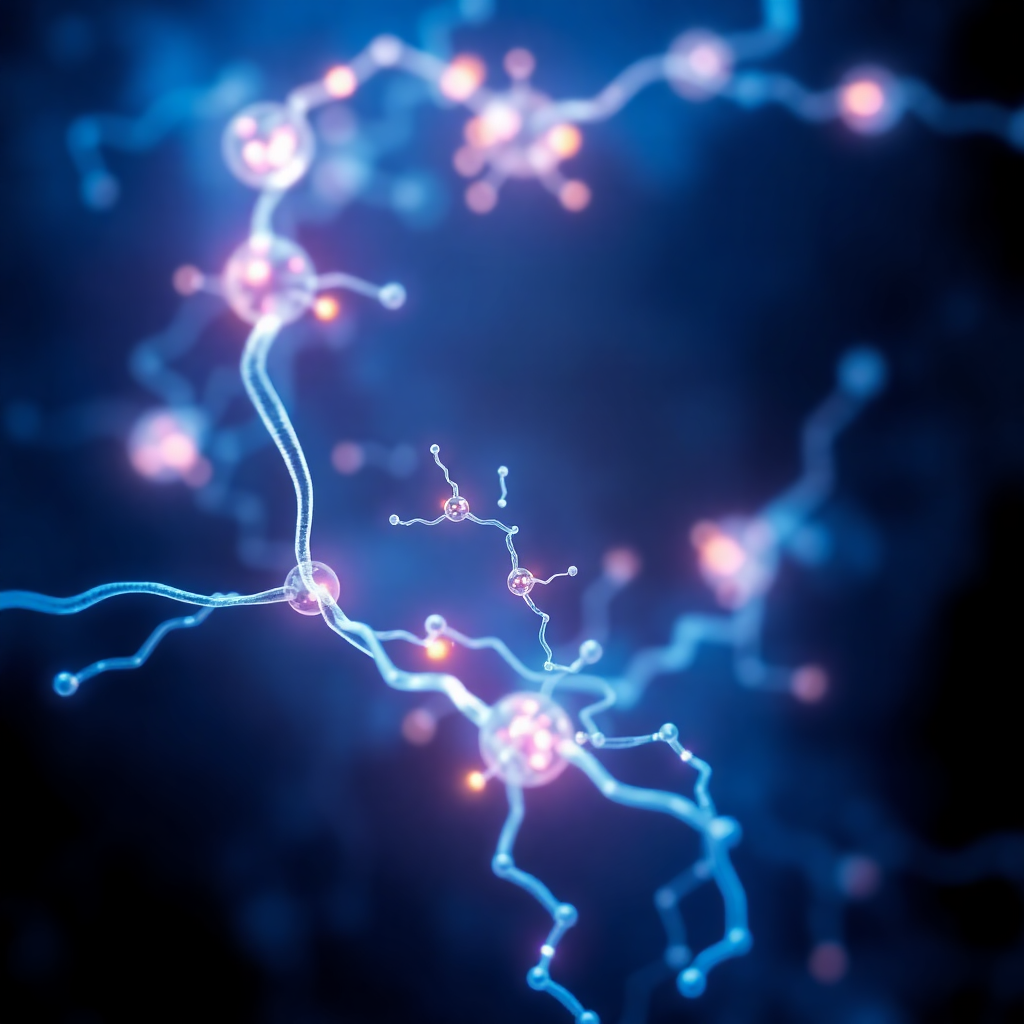
Witness the extraordinary journey of MuseMSCs™ from peripheral circulation to targeted tissue regeneration. A precise, naturally occurring biological process.
STEP-BY-STEP PROCESS
The Journey of a MuseMSCs™
From the moment of administration to complete tissue integration, follow the precise mechanism that makes Muse cell therapy revolutionary.
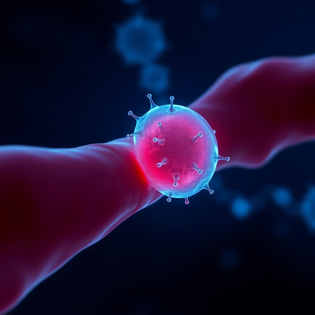
Circulation
Following simple intravenous injection, MuseMSCs™ enter the peripheral blood stream. Due to their uniquely small size (~13μm), they easily bypass the pulmonary trap, circulating freely throughout the body.

Detection & Homing
Damaged tissues emit Sphingosine-1-Phosphate (S1P) distress signals. MuseMSCs™ possess specialized receptors that detect this gradient, allowing them to actively migrate directly to the site of injury.

Phagocytosis
Upon arrival at the damaged tissue, MuseMSCs™ exhibit a unique capability: they phagocytose (engulf and clear) dead or dying cells. This critical step prepares the microenvironment for regeneration.

Differentiation
Finally, guided by the local microenvironmental cues of the cleared tissue, MuseMSCs™ differentiate into the specific cell types required, structurally integrating to restore functional tissue.

BEYOND DIRECT INTEGRATION
Powerful Bystander Effects
While structural integration is their primary function, MuseMSCs™ also exert profound paracrine effects, fundamentally altering the local tissue microenvironment to promote healing.

Immunomodulation
Capable of differentiating into multiple cell types across all three germ layers without the risk of tumor formation inherent to iPS or ES cells.

Angiogenesis
By releasing Vascular Endothelial Growth Factor (VEGF) and other factors, they stimulate the formation of new blood vessels, restoring critical blood supply to ischemic tissues.

Anti-Apoptosis
They provide trophic support to surrounding stressed cells, halting the cascade of programmed cell death (apoptosis) and preserving existing healthy tissue architecture.